Lipoma in Dogs: Signs, Causes & Treatment (Vet Answer)
Updated on

Click to Skip Ahead
Lipomas are a common type of tumor in dogs and are fortunately benign. Lipomas can have a variety of appearances and are diagnosed through cell evaluation under a microscope. Lipomas can cause problems for dogs, however, depending on their location. Continue reading below to learn more about lipomas in dogs.
What Is a Lipoma?
A lipoma is a growth that is made up of fat cells. Generally, lipomas are slowly growing masses that are found underneath the skin. The mass can either be found in the subcutaneous space between the skin and muscle, or sometimes even within the muscle. Rarely a lipoma can be found within the skin or dermis. Infrequently lipomas can be found within the chest or abdomen. Lipomas are benign tumors that do not metastasize or cause illness in pets. However, lipomas can grow in locations that can be problematic for dogs which may necessitate surgical removal.
Infiltrative lipomas are benign but can be locally aggressive, invading surrounding tissue, whereas simple lipomas increase in size and do not disrupt surrounding tissues. Another variant of the lipoma is called myelolipoma. Myelolipomas are mixed with adipose cells and hematopoietic cells, which are the cells responsible for the development of blood cells.

What Are the Signs of a Lipoma?
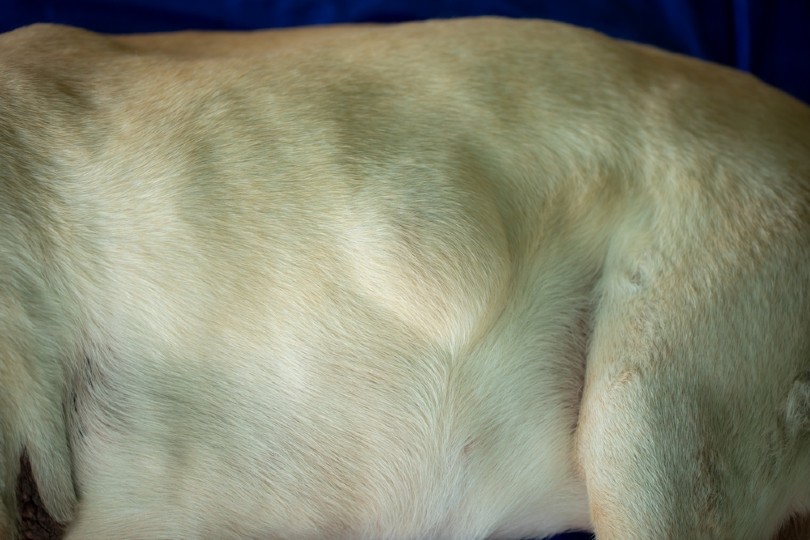
The first sign of a lipoma is a noticeable growth or bump somewhere on the dog’s body. Lipomas tend to form on the trunk of the body but can be found in other locations as well. Lipomas can be firm or soft in feel and can either be attached or freely moveable. An attached lipoma is one that has adhered to another structure like muscle that prevents it from being lifted. If a lipoma occurs within the thorax or abdomen, discomfort and respiratory changes may be noted. Some patients with lipomas may also experience lameness, especially if the mass impacts muscle and range of motion.
Diagnosing a Lipoma
It is important to highlight that a mass cannot be deemed a lipoma without testing. If a dog has previously been diagnosed with a lipoma, it does not mean that all future growths will also be a lipoma. It is critically important to aspirate every mass to correctly identify it. Additionally, masses that undergo a sudden change in appearance should be reevaluated, even if previously deemed benign.
A fine-needle aspiration can be performed by your primary care veterinarian. This involves collecting a cell sample from the mass by gently inserting a small needle into the growth and aspirating cells. Once the cells have been collected in the needle, they are placed on a slide and evaluated under a microscope after the sample has dried and appropriately stained.
During the evaluation of the slide, veterinarians are looking at cells to determine what type of growth is present. Lipomas will have adipose cells present that appear like clear, fluffy, conjoined marshmallows. In some instances, veterinarians may need the assistance of a pathologist to identify cells, and the slide sample may be submitted to an outside laboratory for evaluation.
A biopsy involves collecting a larger cell sample from the mass that occurs once the mass has been removed or with the removal of a small portion of the mass itself. This is a more accurate way of diagnosing a mass, as it involves the evaluation of a larger sample size.
Predispositions for Lipoma Development
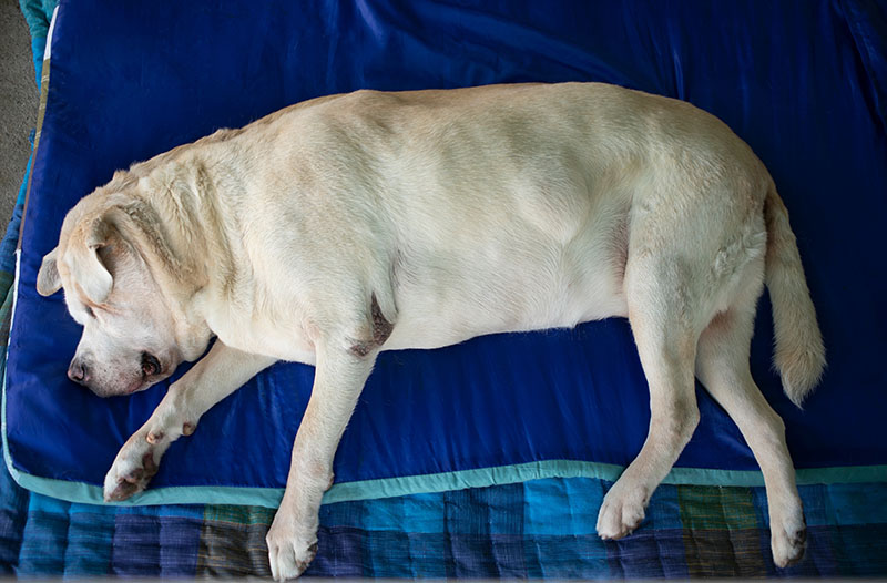
Lipomas are more likely to develop in middle-aged to senior dogs. Overweight dogs are more prone to the development of lipomas. Labrador Retrievers, Beagles, Miniature Schnauzers, and Doberman Pinchers are a few of the breeds predisposed to lipoma development.
Treatment of a Lipoma
Lipomas that are bothersome to the pet or the pet owner can be removed through surgery. It is more helpful to remove the mass when it is small versus when it has been present for a while and increased in size. A smaller mass generally means a less invasive surgery would be necessary.
Infiltrative lipomas may be more difficult to remove and might experience regrowth. Infiltrative lipomas that are not fully excised may benefit from radiation. It is important to note that previously diagnosed lipomas must be closely monitored. If their shape, firmness, or size drastically changes, the mass should be reevaluated and potentially aspirated again.
How Do I Care for a Dog With a Lipoma?

It is important to be cognizant of any changes to the growth. If the mass changes in size, especially if it occurs quickly or if it becomes problematic to your pet, you should schedule an appointment with your veterinarian. Close monitoring of your dog’s mobility is also recommended as often surgical removal of a lipoma is indicated if the mass hinders mobility. Overweight dogs are more prone to the development of lipomas, so keeping your dog trim will reduce their chances of lipoma development. And finally, any new mass that develops should be evaluated by a veterinarian.
FAQs
My Dog Has a Squishy Mass on Their Belly. Is This a Lipoma?
Unfortunately, you cannot diagnose a mass from feel alone. There is a chance that a soft mass may be a lipoma, but the only way of knowing for sure is to evaluate the cells making up the mass.
Are Lipomas Dangerous to My Dog’s Health?
Lipomas themselves are benign; however, depending on their location, they could be problematic to your dog from a comfort standpoint.
Is a Lipoma the Same as a Liposarcoma?
No. A lipoma is a benign growth, whereas a liposarcoma is considered malignant. Liposarcomas are locally invasive and can metastasize to other body regions although very uncommon.
Summary
Lipomas are common growths impacting middle-aged to older dogs. Although they act benignly, close monitoring is recommended. If a growth quickly changes size or is bothersome to the pet, the mass should be reevaluated. Depending on the location of the lipoma, surgery may be recommended to prevent mobility concerns or patient discomfort.
Featured Image Credit: Phatthanit, Shutterstock


